
Blackberries - Your Brain’s New Best Friend
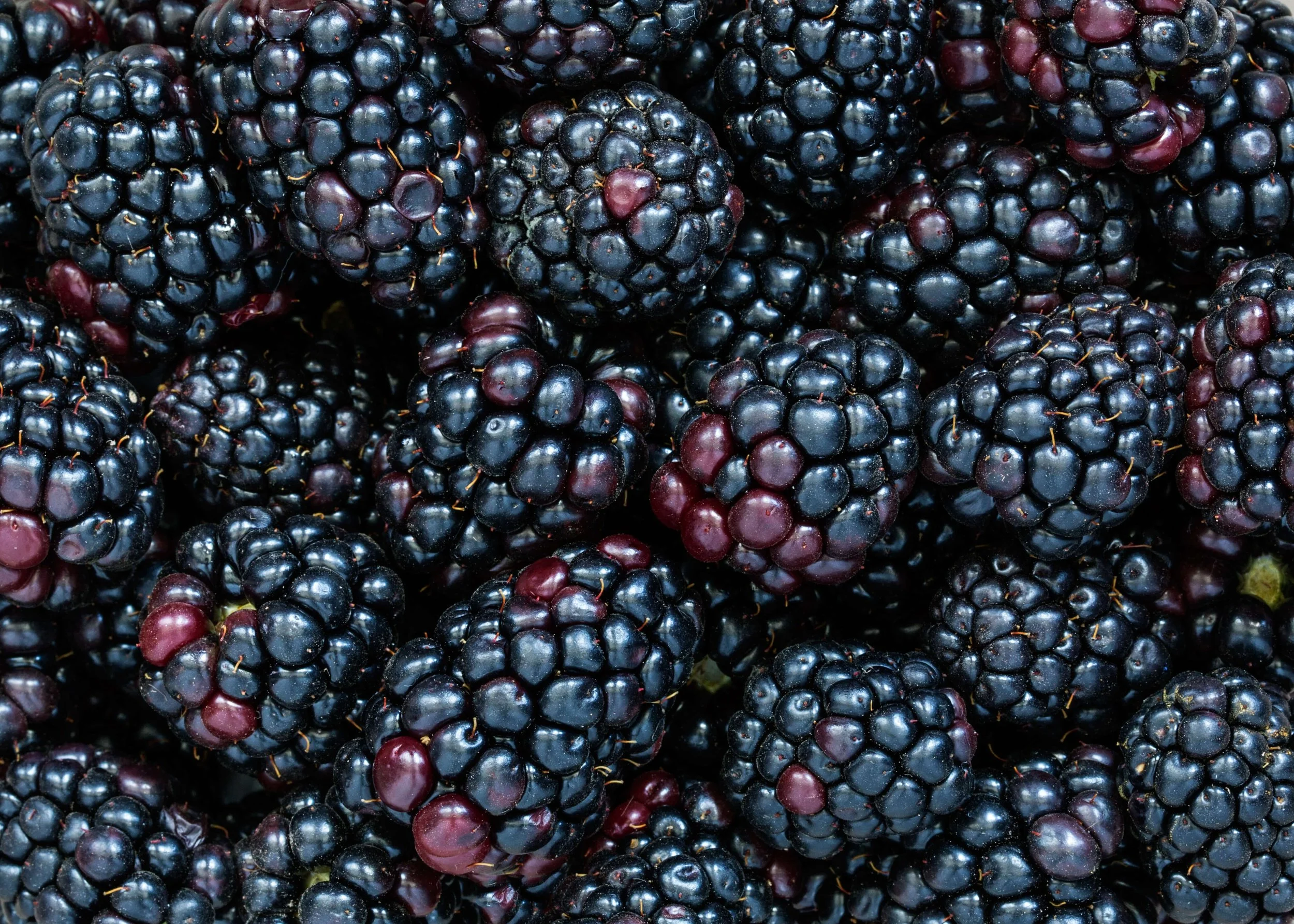
From Celtic folklore, blackberry brambles were prized as a symbol of protection and used for their healing properties. Though the legends differ from science, both recognize the blackberry’s extraordinary capabilities (Driscoll’s 2025). Researchers attribute their potential health benefits to the abundance of polyphenols, which can help reduce neuroinflammation and prevent the onset of neurological diseases.
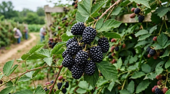
MEET THE BLACKBERRY
Blackberries, scientifically known as Rubis fructicosis, are perennial plants closely related to raspberries. What’s the difference? When you pick a raspberry, it slips off its white core, leaving it hollow inside. Blackberries, on the other hand, retain their central “torus” core when picked (Driscoll’s 2025).
Blackberries originally came from temperate regions in the Northern Hemisphere and were distributed worldwide through European colonization (Grant, 2022). Along the way, they became more than just a source of food; in ancient Greece, they were used to cure gout and sore throats. During the Civil War, they were thought to relieve dysentery and stomach ulcers (Driscoll’s 2025). Today, while they’re perhaps best known for making a mean cobbler, research suggests there’s more to these berries than just their flavor.
AN INTRODUCTION TO POLYPHENOLS
Blackberries are rich in polyphenols, which are a class of chemical compounds found in many plants (McDermott & Rose-Francis, 2026). There are more than eight thousand different types of currently identified polyphenols (Nock, 2021). Plants use polyphenols to support healthy development. These compounds make sure that plants can absorb the sunlight they need to grow, ultimately protecting the plant (Foods Rich in Polyphenols — and Why They’re Important 2023). But humans don’t photosynthesize! Bummer. But we can use polyphenols differently. Polyphenols have two main benefits for the body: they’re antioxidants and anti-inflammatory (Nock, 2021). Let’s break it down even further.
Because there are thousands of different polyphenols, scientists group them into four major categories: flavonoids, phenolic acids, lignans and stilbenes. So… what’s the difference? Well, the differences lie in their chemical structures.
All polyphenols contain one or more “phenol units.” These units consist of a benzene ring bonded to another group of elements called the hydroxyl group. In plants, however, these units rarely exist on their own. Instead, they are found in “conjugated forms,” meaning a sugar molecule usually attaches onto the hydroxyl group or directly to the aromatic ring (Ciupei et al., 2024). This seemingly small difference affects the compound’s stability, solubility and biological activity.
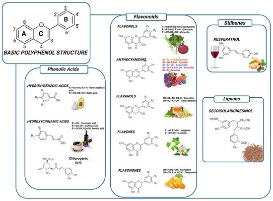
The small structural differences determine how a compound is classified. Let’s take a closer look at flavonoids, which are the largest and most extensively studied group. Flavonoids generally contain two six carbon aromatic rings connected by a three carbon “bridge” to another ring. The placement of the hydroxyl group and sugars give rise to many subclasses of flavonoids (Ciupei et al., 2024).
Anthocyanins are one such subclass, and they can be distinguished by their “flavylium ion structure.” What does that mean? It means that the three-carbon bridge also contains a positively charged oxygen atom. This structure, with the two rings and modified bridge is called the flavylium ion (Banji et al., 2022). Because of this unique structure, anthocyanins absorb light differently than other flavonoids, resulting in the deep blue color seen in blackberries (Robinson et al., 2020).
WHAT IS NEUROINFLAMMATION?
Okay. So, let’s put this all together. How can anthocyanins help reduce inflammation? You might have heard that term passed around a lot: “inflammation.” Inflammation is one of the body’s natural defense mechanisms. When you get a cut or injure a muscle, your immune system initiates a response to begin the healing process. This short-term response, called acute inflammation, is your body’s way of saying “Something’s wrong– let’s fix it.” Once the problem is handled, the inflammatory response subsides (Inflammation, 2024) . While acute inflammation is healthy, chronic inflammation does more harm than good. When the immune system is exposed to persistent stress (like poor diet or toxins), the inflammatory response lingers. Instead of helping to heal healthy tissue, it malfunctions and begins attacking healthy tissues and organs (Gartry & Kordrostami, 2026).
To grasp this concept, we need to understand glutamate. Glutamate is the brain’s primary neurotransmitter, another type of chemical messenger that allows neurons to communicate with one another. Every thought, memory and movement depends on glutamate (Magdaleno Roman & Chapa González, 2024). You might have seen photos of neurons, depicted in a kind of “web.” At each point where two neurons meet, they communicate across a small gap called the synapse. When the “presynaptic neuron” wants to send a message, it releases glutamate into the synapse (The Editors of Encyclopedia Britannica, 2026). The glutamate then binds to receptors on the “postsynaptic neuron,” and almost immediately afterward, a specialized cell called an astrocyte removes excess glutamate from the synapse.
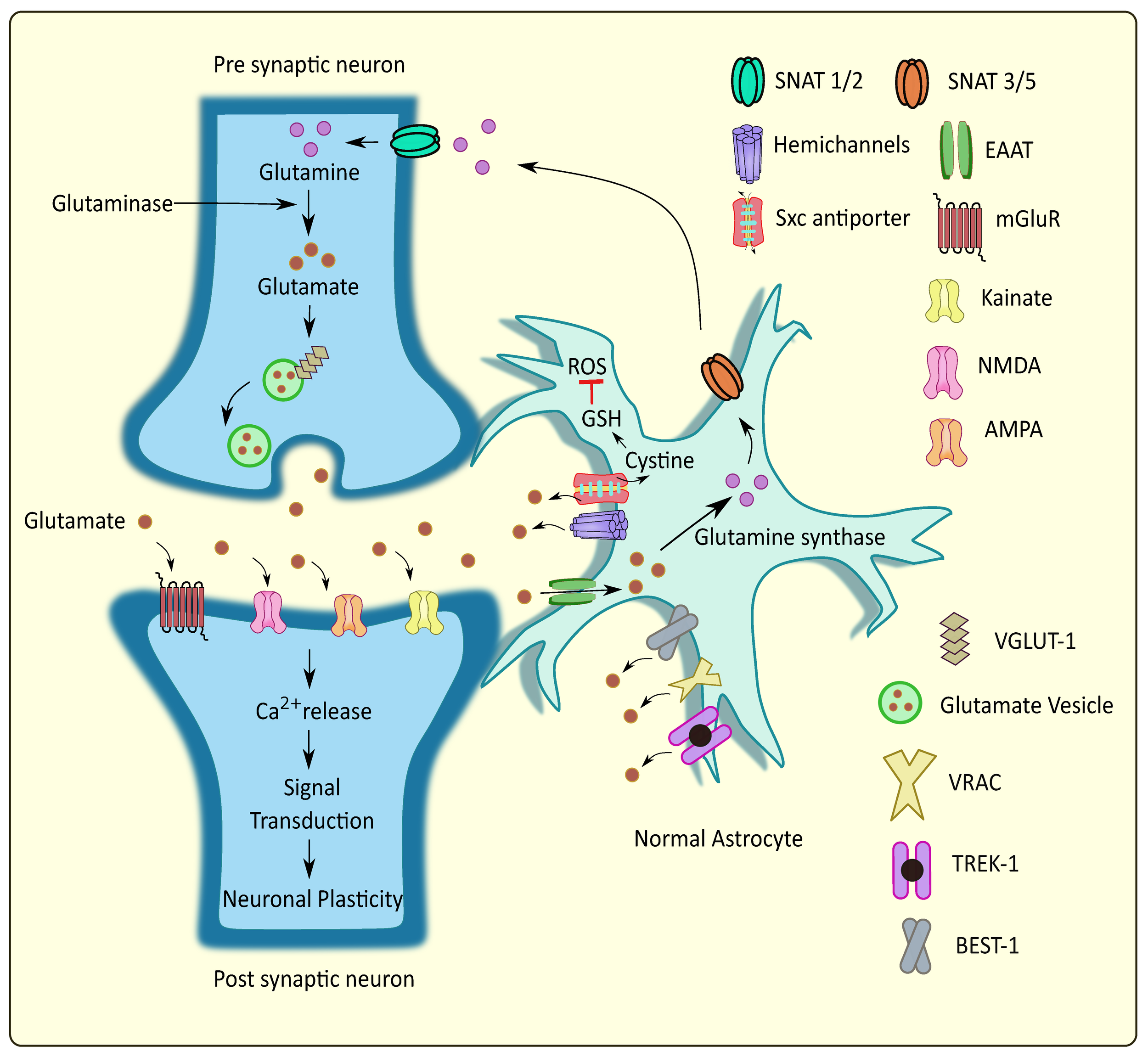
This constant cycle occurs continuously, allowing messages to be sent throughout the brain in milliseconds. So… what would happen if glutamate wasn’t removed? Well, when glutamate is released, it bonds to receptors called “ion channels.” The channels remain closed until glutamate unlocks them, allowing both sodium and calcium to travel into the next neuron (Carvalho et al., 2014). Sodium helps carry the electrical signal onto the next neuron, while calcium tells the neuron when to release signals (Magdaleno Roman & Chapa González, 2024). However, when chronic neuroinflammation disrupts the balance, astrocytes go into “damage control mode” and glutamate begins to accumulate outside neurons. Because the glutamate remains bonded to the receptors, calcium floods into the neuron, causing an overload. This is known as glutamate excitotoxicity, and it overstimulates the cell, and triggers enzymes that causes the cell to consume its own organelles until eventual death (Zhong et al., 2023). It is a major underlying component in Alzheimer’s, Huntington’s, Parkinson’s and Multiple Sclerosis (Banji et al., 2022).
In the brain, inflammation doesn’t look the same as in the rest of the body. Instead, inflammation affects how cells behave and communicate. The brain’s resident immune cells, microglia, shift from their monitoring state and become activated. Once activated, they release proteins called cytokines, which are essentially “chemical messengers” that tell other cells how to respond (Banji et al., 2022). Cytokines diffuse through the brain until they encounter cells with the right receptors. There are many types of receptors, and many types of cytokines. Think of cytokines as keys, and the receptors as locks. They fit together, but each cytokine can only “unlock” certain intracellular pathways. When the cytokine bonds, it sends a signal into that cell that changes its behavior (Banji et al., 2022). While it varies cytokine to cytokine, the signal may cause the cell to release other cytokines, activate other immune cells, express inflammatory proteins, or alter the way it communicates with other cells (Banji et al.). Let’s look at an example:
Imagine someone experiences a fall and gets a concussion. The impact damages some neurons, and the injured cells release danger signals (damage-associated molecule patterns). Microglia detect these danger signals, become activated, and release cytokines like TNF-ɑ or IL-1β (Interleukin-1 Beta), which diffuse through the brain and bind to nearby receptors (Li et al., 2021). This acute inflammatory response is usually temporary. Once the debris is cleared and the damaged tissue is repaired, cytokine production decreases, microglia return to their monitoring state and the brain goes back to “normal” (Banji et al.).
Now, let’s say you’re a wrestler. You have just suffered your tenth concussion. With repeated trauma, the brain doesn’t have time to fully resolve the injury before another injury occurs. In this way, microglia remain chronically activated, releasing cytokines continuously and keeping the brain in an inflammatory state. When microglia remain active like this, signalling can interfere with regular brain function. How?
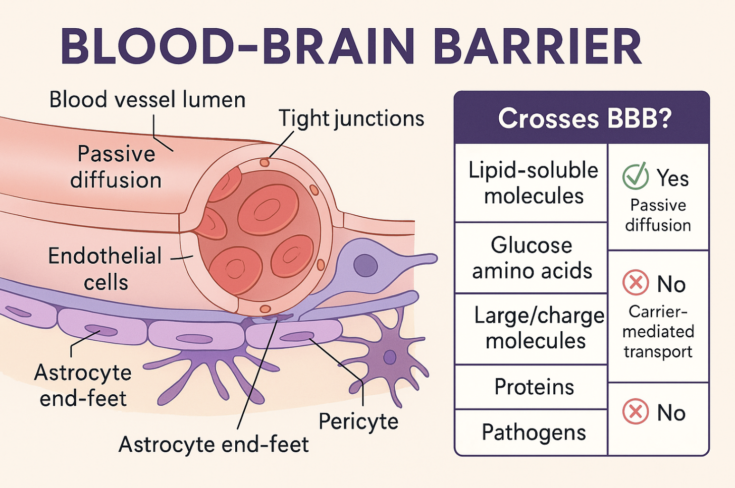
Your brain is so important, it has its own barrier to prevent harmful substances! Cool, right? The blood-brain barrier is a layer of endothelial cells joined by junction protein, sort of acting like mortar between bricks. It forms a membrane that prevents harmful substances from entering the brain while still allowing essential nutrients through (Daneman & Prat, 2015). Not today, pathogens!
However when cytokines are released during a neuroinflammatory response, they can also bind to cells on the blood-brain barrier and cause them to break down the proteins that usually seal neighboring cells together (Daneman & Prat). A perfectly sealed blood-brain barrier would make it difficult for repair molecules to reach the damaged area, so cytokines can make it temporarily more permeable. Issues arise if inflammation becomes chronic, and the barrier can become permanently “leaky” with gaps (Daneman & Prat).
BACK TO BLACKBERRIES
So where do blackberries fit into all of this? While they are certainly not a cure for neurodegenerative disease, new experimental research suggests that anthocyanins in blackberries can help interrupt some of the inflammatory pathways that drive chronic neuroinflammation (Banji et al., 2022). When microglia become activated and move to clear debris, they naturally produce reactive oxygen species (ROS) as part of their defense system (Banji et al.). In normal amounts, ROS helps destroy damaged material, but when microglia stay activated for too long, ROS builds up in excess. This acts as a signal that keeps inflammatory pathways switched on, making microglia even more reactive. A vicious cycle is initiated: microglia produce ROS, ROS reinforce inflammatory signaling, and that signaling keeps microglia in an activated state (Banji et al.).
Anthocyanins target how ROS are produced and used inside microglia. When ROS levels rise, they act as internal signals that push inflammatory pathways into a more active state, causing an uptake in cytokine release (Zhong et al., 2023; Banji et al., 2022). Anthocyanins activate the cell’s antioxidant pathways which lower the ROS levels in a cell. With less ROS available to trigger inflammatory reactions, microglia produce less cytokines and are less likely to remain in a prolonged activated state (Banji et al.). By reducing cytokine release, anthocyanins support healthy astrocyte activity. In this way, they can continue clearing glutamate from the synapse normally, preventing excitotoxicity and neuron death (Li et al., 2021). Although anthocyanins aren’t exactly an “off-switch” for inflammation, they can weaken one of the key signals that keeps the inflammatory cycle running.

PIECING IT ALL TOGETHER
It is important to note that much of what is known about anthocyanins and its implications for neuroinflammation comes from experimental research rather than clinical treatment. Many of the mechanisms described have been observed primarily in cell cultures and animal studies, where the conditions are carefully controlled (Banji et al., 2022; Li et al., 2021). These models are powerful for revealing how biological systems work, but to understand the full scope of its implications, one would need to fully replicate the complexity of a living human brain. This is a difficult undertaking because the brain cannot be directly manipulated in a living person due to ethical limits in clinical research. Researchers cannot intentionally induce brain injury or alter neural pathways to observe outcomes in real time, so human studies rely on indirect measures such as imaging and overall cognitive performance. While the evidence suggests meaningful neuroprotective potential, these findings are still being tested and refined (Banji et al., 2022). Still, despite these limitations, the repeated patterns seen across experimental models offer a compelling starting point for understanding how small dietary compounds may interact with the brain’s systems.
Every study adds another piece to a much larger picture of how food, molecules, and brain function are connected in ways we are only beginning to understand. Something as simple as a berry, once known only through folklore and tradition, is now part of a larger story: the brain and its mysteries. It is now entangled in a network of over eight billion neurons, one hundred trillion synaptic connections and everything that makes you, “you.”
Perhaps even greater, every study only contributes to my burning desire to eat lots of cobble and consume blackberries by the pound. After all, blackberry cobbler is basically brain research in dessert form, and I intend to continue my investigations accordingly.
